Our skin develops various growths and changes that are normal as we mature. Many of these skin changes are benign and represent normal aging, sometimes due to intrinsic factors (i.e., genetics & natural age progression changes) and/or extrinsic factors (i.e., sun exposure). The purpose of this article is to provide you with basic background information for better understanding. It is important to have a qualified skin expert (Board-certified Dermatologists) confirm these growths are benign (non-cancerous) and not malignant (cancerous).
Benign skin growths do not require removal or treatment. Many people desire to have them removed, and there are various techniques often yielding excellent cosmetic outcomes. The procedures are almost always a non-covered benefit of medical insurance.
SOLAR LENTIGO (also known as SUN SPOT, LIVER SPOT)
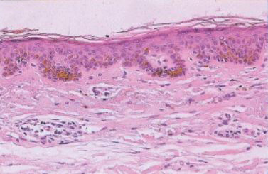
A solar lentigo (lentigines, plural) is a tan, brown or dark brown flat discoloration of the skin usually 2 mm to 2 cm in size, most commonly appearing on sun-exposed areas of the body, including the face, neck, chest, shoulders, arms, hands, and legs. They represent excess pigmentation in the skin caused by ultraviolet light exposure over time.
They do not require treatment; however, intense pulsed light and/or fractional resurfacing lasers with some topical therapies yield excellent results.
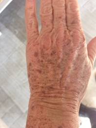
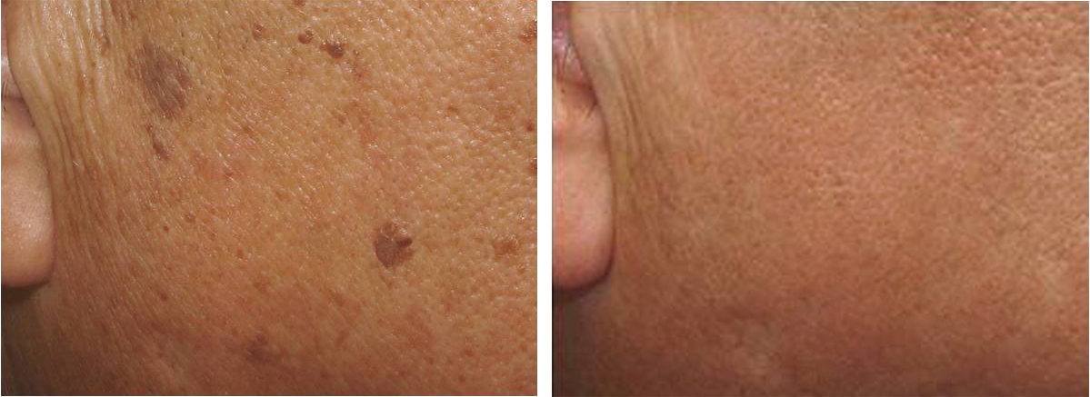
Example of Lentigo & Treatment Results
(Photo Above: sunspots before treatment)
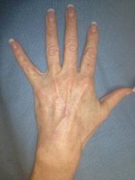
(Photo Above: same hand after one IPL treatment)
Dr. Liu features on the Doctors Show Treating Solar Lentigos with Laser Therapy
(Video Above: Live Demonstration of Patient Above)
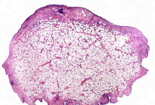
Lentigo Microscopic Example
SEBORRHEIC KERATOSIS (also known asWarty Keratosis, Stucco Keratosis, Barnacles)
Seborrheic keratoses are benign skin growths of the outer layer of skin (epidermis), often described as “waxy”, “stuck-on”, “warty”, or “barnacles.” They may be few or numerous in number. The color is often brown, but can be skin-colored or black. They can appear on almost any part of the body including the face, scalp, torso, extremities, and genitals. Seborrheic keratoses may exhibit more than one color and grow quickly, prompting patients to visit the Dermatologist.
Seborrheic keratoses can be successfully removed using a variety of techniques, including cryotherapy, electrodessication, surgery, and/or laser.
Dr Liu explaining Seborrheic Keratosis on the Doctors Show
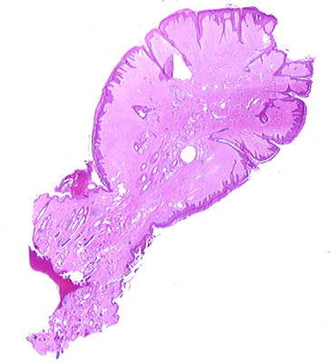
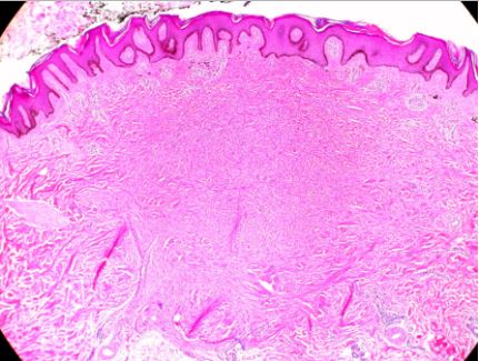
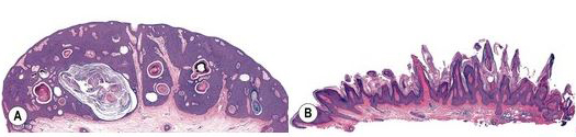
Seborrheic Keratosis Microscopic Example
(Left above is an example of the hyperplastic variant; Right above is example of warty variant)
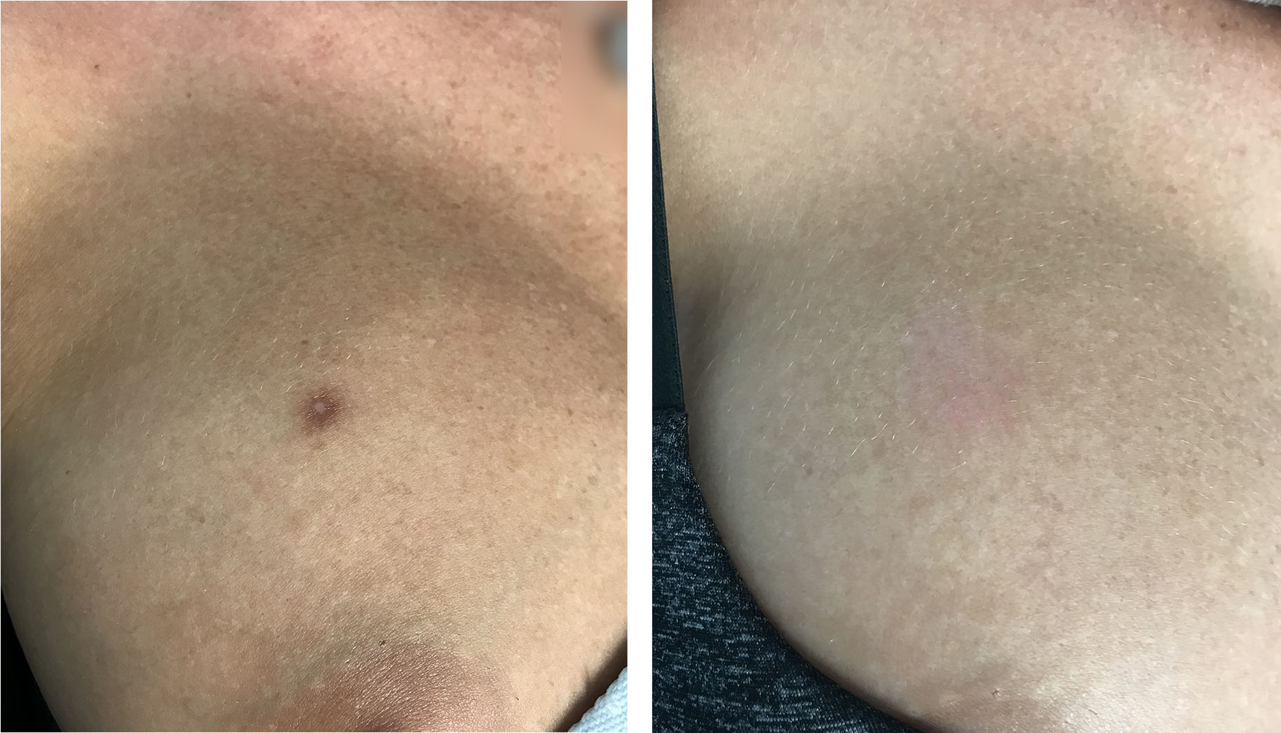
Example of Treatment Results of Seborrheic Keratosis
Seborrheic Keratosis & Sun Spot Treated with Fractional Laser & Intense Pulsed Light
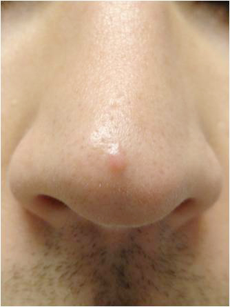
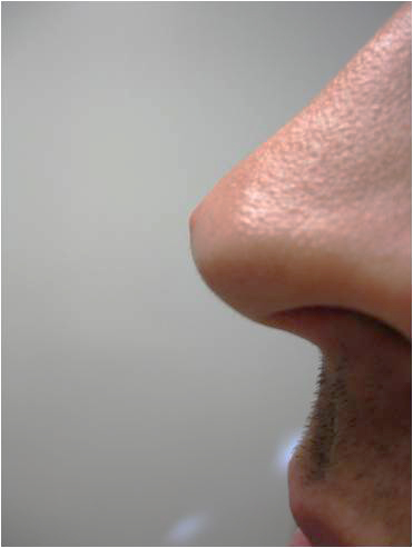
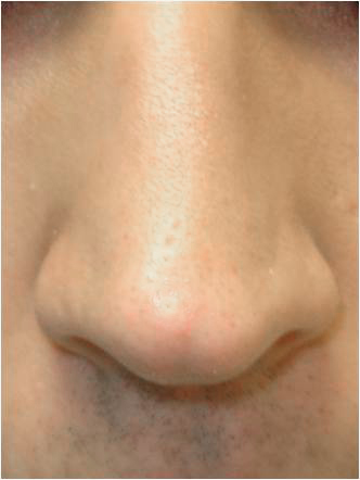
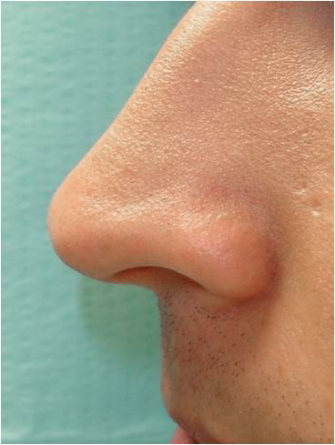
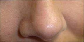
FIBROUS PAPULE (also known as ANGIOFIBROMA)
Benign growth composed of collagen fibrous tissue and blood vessels. They range from white-skin colored to red, often with a smooth surface appearance. They occurs most commonly on the face, particularly on the tip or side of the nose;
Cosmetic shave contouring, electrocautery (electric heated needle tip) and/or laser treatments can successfully remove these growths.
HEMANGIOMA (also known as CHERRY ANGIOMA)
Adult onset hemangiomas are benign clusters of dilated blood vessels. They range from bright red to purple, often with a smooth surface appearance. They occurs most commonly on the torso; however, a hemangioma can develop anywhere on the body. Individuals may develop a few to hundreds of them.
Electrocautery (electric heated needle tip) or laser treatments can successfully remove these spots.
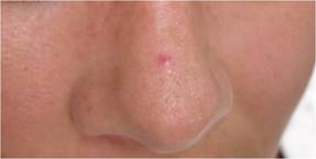
Example of Hemangioma & Treatment Results
(Photo Above: common location of cherry angioma on the nose)
(Photo Above: after one treatment with intense pulsed light)
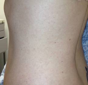
Example of Hemangiomas on Torso
(Photo Above: hundreds of cherry angiomas on torso)
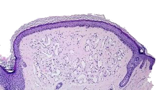
Example of Hemangioma under Microscope
Collection of small benign extra blood vessels just underneath the skin surface
SKIN TAGS (ACROCORDONS)
Skin tags are soft benign skin growths that hang off the skin surface. They are often skin-colored but can appear pink, tan, or brown. They most commonly form in areas of friction, such as the neck, armpits, and groin. They are relatively frequent around the eyelids as well.
Skin tags can be successfully treated using snip removal, electrodessication, and/or cryosurgery.
Clinical Example of Skin Tag
(Photo Above: Skin tags developing on armpit area)
Skin Tag Under Microscope
SEBACEOUS HYPERPLASIA
Sebaceous hyperplasia growths are smooth or waxy, small white or yellow bumps on the face commonly mistaken for whitehead pimples. They are frequently unsuccessfully extracted during facials. These benign growths are enlarged clusters of normal oil glands in the skin, most commonly appearing in the 3rd or 4th decade of life and can increase in number and size over time.
Sebaceous hyperplasia can be successfully removed using electrodessication and/or laser.
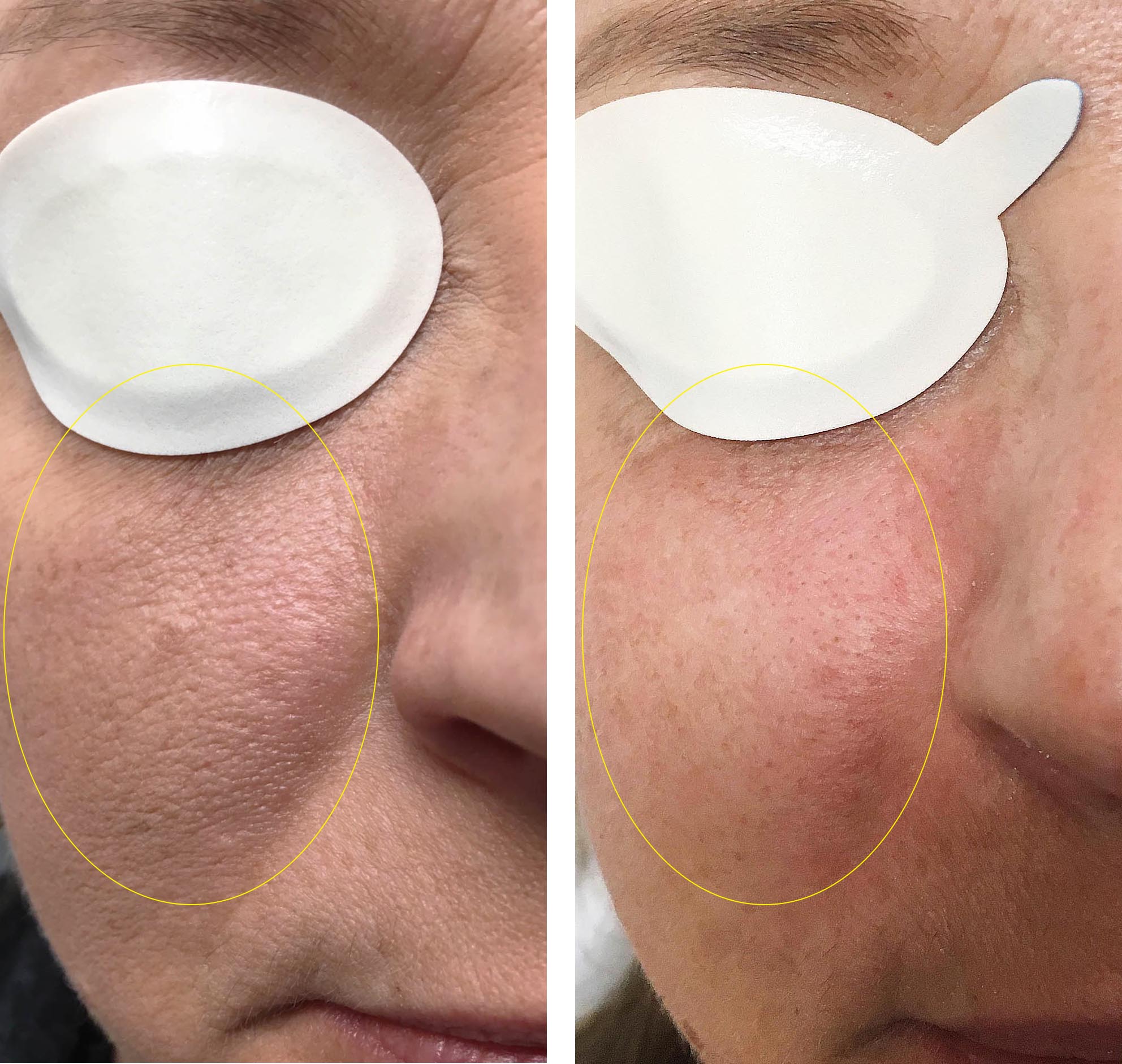
Clinical Example of Sebaceous Hyperplasia
(Photo Above: Sebaceous hyperplasia on nose and cheeks)
(Photo Above: Sebaceous hyperplasia on cheek treated using laser)
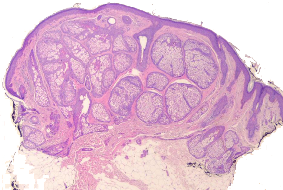
Sebaceous Hyperplasia Under Microscope
Extra benign oil glands under surface of skin
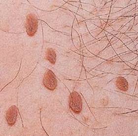
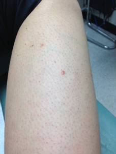
DERMATOFIBROMA
Dermatofibromas are often solitary growths that appear over a few weeks. The color can range from pink-red, to brown often shaped in a dome and firm to the touch. They can sometimes exhibit a feature called the “dimple sign” where the growth dimples into the skin when pinched. They occur most commonly on the legs of women, but also on the arms & shoulders of both men and women. Dermatofibromas represent a scar-like reactive process that may be induced by a trauma, such as an insect bite. They often grow to a certain size and stay.
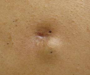
EPIDERMAL INCLUSION CYST (also known as SEBACEOUS CYST, FOLLICULAR HAIR CYST)
An epidermal inclusion cyst is a benign growth composed of an incomplete hair follicle-oil gland lining, filled with keratin. The produced keratin becomes compact into a cheese-like material. They sometimes have a small opening from which the malodorous keratin can be expressed or no surface opening, which appears as a smooth bump underneath the skin surface. They often grow to a certain size then stay. If the cyst lining ruptures, a boil or abscess can form.
Surgery is the preferred technique for removing cysts.
(Photo Above: Epidermal Inclusion Cyst treated by incision & drainage resulting in scar & recurrence)
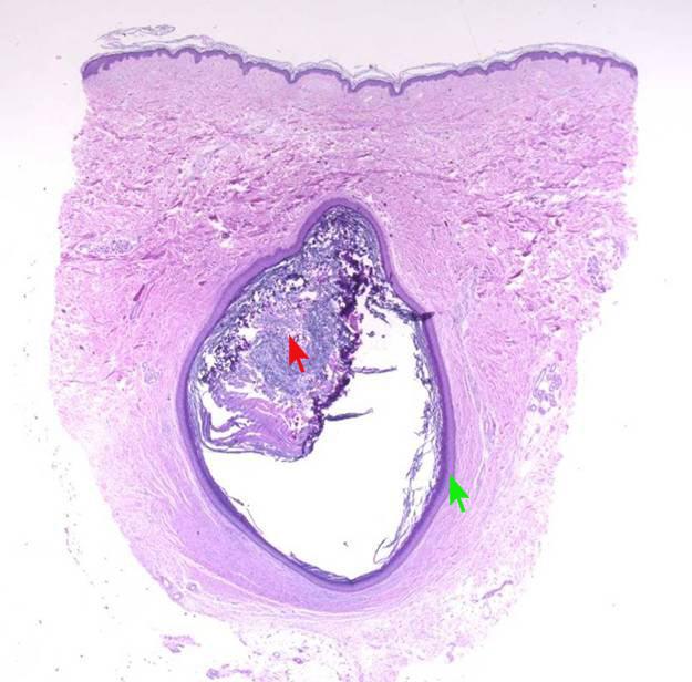
Epidermal Inclusion Cyst Under Microscope
MILIA
Milia can be thought of as miniature epidermoid cysts. They frequently occur around the eyelids of young women, but can appear on other parts of the face and body on men or women.
Milia can be effectively removed using extraction, electrodessication, and/or laser.
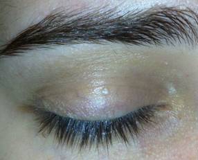
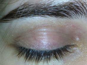
Example of Milia & Treatment Results
(Photo Above: Milia on eyelid before treatment)
(Photo Above: Same eyelid after electrodessication)
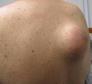
LIPOMA
A lipoma is a benign fatty tumor that lies deep in the skin and appears as a soft lump. Occasionally, lipomas are tender to the touch but usually do not cause other symptoms. Lipomas may be small or quite large and develop in adults as single or multiple lesions, sometimes hereditary.
Surgery or liposuction yield excellent results.
Dr Liu Featured on the Doctors Show Surgically Removing a Lipoma


 Collection of small benign extra blood vessels just underneath the skin surface